Dinoflagellates

24 July, 2009


In my micropaleontological studies in the 1980’s I dealt with peridinioid dinoflagellates, as a morphologically defined group, from the Upper Cretaceous (Maastrichtian) of Trelleborg at the southwestern part of Scania (Skåne), southern Sweden. My intention was to contribute to the taxonomy of this group by describing the forms present, recording their dimensions, and analysing the relations in shape between endocyst and pericyst.

Fossil dinoflagellates that appear morphologically related to modern Peridinium and Protoperidinium were treated as peridinioid dinoflagellate cysts. Several types, styles, and shape variants of archeopyles were recognized in this group.

In peridiniacean genera as a rule the archeopyle corresponds to the second anterior intercalary paraplate (2a) and the inferred paratabulation is of the hexastyle.

Genera which reveal indications of a quadrastyle paratabulation or have intercalary archeopyles corresponding to more than one paraplate (e.g. Trithyrodinium) or combination archeopyles corresponding to paraplates of different series (e.g. Palaeoperidinium) were not included in the peridiniacean genera, but regarded as peridinioid.

In papers on dinoflagellates detailed recordings of dimensions of the material studied and calculations on those data were rarely presented. Before 1980 I found only three studies that used morphometry:

  • Regression analysis was used to study the relation between horns and cyst body
  • The distribution of length/width ratio was used to study the interspecific variation and taxonomy of Alterbia
  • Dimensions of length, width, and length of horns and spines were graphically presented in one paper

I have treated ten species of acid resistant peridinioid dinoflagellates of Deflandrea, Isabelidinium, Palaeoperidinium, Phelodinium, Subtilisphaera, Svalbardella, and Trithyrodinium from this Upper Cretaceous (Maastrichtian) material from southern Sweden.

Taxonomical conclusions

  • The discrimination between Isabelidinium Lentin & Williams (1977) and Eurydinium Stover & Evitt (1978) according to the diagnoses, by means of length/width ratio of the endocyst is not justified in my studies. This ratio displays a normal distribution which includes values which originally have been considered critical for the discrimination of two separate genera.
  • The features discriminating Isabelidinium Lentin & Williams (1977) and Chatangiella Vozzhennikova (1967) are not clearly indicated in the original descriptions. Most of the diagnostic differences may be modifications due to environmentally controlled factors. In preserved material those morphologic features seem to display ranges which include both genera. I have published the new combination Isabelidinium armatum (Cookson & Eisenack) Lindgren.
  • Lejeunia kozlowskii Górka (1963) is a cornucavate cyst referable to Phelodinium Stover & Evitt and different from Lejeunecysta tricuspis (Wetzel) Artzner & Dörhöfer (1978). I have published the new combination Phelodinium kozlowskii (Górka) Lindgren.
  • Palaeocystodinium Alberti (1961) is a later synonym of Svalbardella Manum (1960). So is Cystodiniopsis Vozzhennikova (1963) which name however is illegitimate; the designation of a new type species is contrary to the ICBN.
  • Ceratiopsis Vozzhennikova (1967) and Pentagonum Vozzhennikova (1967) as names of dinoflagellate genera are illegitimate and must be rejected, as they are later homonyms of Ceratiopsis De Wildeman (1896) and Pentagonium Schauer (1843), which were validly published for a genus of fungi and an asclepiadacean genus, respectively.

References

Lindgren, S., 1984.
Acid resistant peridinioid dinoflagellates from the Maastrichtian of Trelleborg, southern Sweden. (Summary in Russian.) Stockholm Contrib. Geol., 39(6): 145—201. Stockholm. ISSN 0585-3532. ISBN 91-22-00519-6. — Buy at the lowest prices among books in Sweden.
Lindgren, S., 1985.
Nomenclatural notes on fossil peridinioid dinoflagellates. Taxon, 34 (4): 670—671. Utrecht. ISSN 0040-0262.

Web Links

Dinoflagellates; by Matthew Olney, University College, London.

Dinoflagellates; by Mona Hoppenrath and Juan F. Saldarriaga, Forschungsinstitut Senckenberg, Germany, and University of British Columbia, Canada

Introduction to the Dinoflagellata; by Graham Williams & Andrew MacRae, Museum of Paleontology, University of California, Berkeley.

Peridinium; by Charles J. O’Kelly, Protist Image Database.

Graham Williams, Dartmouth, 1996

Charles Downie, Sheffield, 1923—1999

Bill Sarjeant, Saskatoon, 1935—2002; also in The Encyclopedia of Saskatchewan.

Lewis Stover, Kerrville, 1925—1993

Tamara Vozzhennikova, Novosibirsk, 1914—2000


On the illegitimate status of genus Ceratiopsis

24 July, 2009
 In May 1992 I was contacted by a collaborator of the Index Nominum Genericorum in Utrecht. She wrote:

”Recently I met your article in Taxon 34: 670—671, 1985, stating that Ceratiopsis was not validly published by Vozzhennikova 1963: 181. As far as I can see, however, in 1963: 181, there is one species only, thus the name was validly published under art. 42. Please tell me from which you drew the conclusion that the genus comprised three species.”

My answer was simple:

“My conclusion that the genus Ceratiopsis comprised three species is drawn simply from the original text by Tamara Vozzhennikova, which states in Russian “ТРИ ВИДА” (tri vida) = three species.”

The reply from the ING collaborator was a little surprising:

”I would be inclined to say that Note 1 to Art. 42 applies: ‘… the author may indicate that other species are attributable to the genus.’ I suppose that this has been [another ING collaborator’s] interpretation as well, when he made ING’s entry for Ceratiopsis.”

Surprising, because the Article 42 has no notes at all!

My Taxon article was published in 1985 and I had to use the Code edition that was really published and effective at that time. That code article 42 deals with publication of the name of a monotypic new genus. As stated in Vozzhennikova’s original paper the genus comprised three species (Russian: “tri vida”) so it evidently was not monospecific.

By changing the rules of the Code it is possible for the international congresses to keep scientists of the world busy working with reinterpretations of previous results. I do not want to join such a play.

On the internet there are links to two references (link 1 and link 2) to Lentin & Williams, 1987. I have not seen that article.

However, my Taxon article 1985 have another point concerning the status of genus Ceratiospsis:

Ceratiopsis Vozzhennikova 1967 as name of a dinoflagellate genus is illegitimate and must be rejected, as it is a later homonym of Ceratiopsis De Wildeman 1896, which was validly published for a genus of fungi (ICBN 1978, Article 64).


References

De Wildeman, E., 1896.
Quelques notes sur la nomenclature générique des champignons. Bull. Séances Soc. Belge Microsc. 22 (6): 108—119. Bruxelles.
International code of botanical nomenclature, adopted by the twelfth international botanical congress, Leningrad, July 1975.
Utrecht, 1978: Bohn, Scheltema & Holkema. ISBN 90-313-0332-1.
Lentin, J.K. & Williams, G.L. 1987.
Status of the fossil dinoflagellate genera Ceratiopsis Vozzhennikova 1963 and Cerodinium Vozzhennikova 1963 emend. Palynology, 11: 113—116.
Lindgren, S., 1984.
Acid resistant peridinioid dinoflagellates from the Maastrichtian of Trelleborg, southern Sweden. (Summary in Russian.) Stockholm Contrib. Geol., 39(6): 145—201. Stockholm. ISSN 0585-3532. ISBN 91-22-00519-6. — Buy at the lowest prices among books in Sweden.
Lindgren, S., 1985.
Nomenclatural notes on fossil peridinioid dinoflagellates. Taxon, 34 (4): 670—671. Utrecht. ISSN 0040-0262.
Vozzhennikova, T.F., 1963.
Tip Pyrrophyta. Pirrofitovye vodorosli (in Russian, Algae of Pyrrophyta). In: Osnovy paleontologii. Spravochnik diya paleontologov i geologov SSSR, tom (14) “Vodorosli …” p. 171—195. Izd. Akad. Nauk SSSR, Moskva, 698 p.


Morphometry of peridinioid dinoflagellates

24 July, 2009

In peridinioid dinoflagellates of the Maastrichtian (Upper Cretaceous) of Trelleborg (Scania, southern Sweden) analyses of the length/width ratio of the pericysts and the endocysts of different species also indicate that the variation of the pericysts generally is greater than that of the endocysts. The endocysts display a less elongated shape which is fairly constant within a species. The specific mean values of the length/width ratio of endocysts range between 0.8 and 1.4 with the median value = 1.0, while the specific mean values for the pericysts range from 1.3 to 4.5, with the median value = 2.1

In these peridinioid dinoflagellates the restricted variation in size and shape of the endocysts indicates that the endophragm was developed under strong genetic control inside the motile, vegetative theca. The pericysts, however, display a wide morphologic variation which may be the result of influences by environmentally controlled factors during the development of the periphragm. This development may have taken place also on the outer side of the motile theca, or even after its decay.

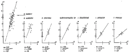
Analyses of the Trelleborg material of the length/width ratio of pericysts and endocysts by reduced major axis in scatter diagrams indicate that within a genus the slopes of the regression lines are close to each other within a genus with statistically significant correlation coefficients. Thus the slope of the regression lines may be used for taxonomical purposes.

See also >> Dinoflagellater: mätningar och klassifikation (in Swedish only)


Dinoflagellater: mätningar och klassifikation

24 July, 2009


Dinoflagellater är en grupp encelliga, mikroskopiska alger som kan variera kraftigt till utseendet. Dinoflagellaterna är kända från silur men förekommer främst från mellersta mesozoikum. De är i nutiden en viktig del av havens plankton men lever även i sötvatten.Dinoflagellaternas livscykel har två stadier: det vegetativa stadiet med en theca och ett vilstadium med en cysta. I thecastadiet är cellen aktivt rörlig och omges av ett cellulosaskal. I vilstadiet bildar cellen som skydd mot ogynnsamma miljöförhållanden ett hölje innanför thecan och omkring plasman. Cystan är orörlig.
Hos levande dinoflagellater är det främst theca-stadiet som studerats. Inom paleontologin är det vilstadiet som är intressant eftersom endast cystorna bevaras som fossil.
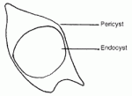
Eftersom cystan bildas innanför thecans vägg och eftersom båda är kontrollerade av samma genotyp är det naturligt att de kan uppvisa betydande likheter så att cystan motsvarar thecans morfologi. Cystornas form kan variera kraftigt. De kan vara sfäriska och släta, men de har ofta olika slags utskott. Flera former är tillplattade. Peridinioida dinoflagellater är tillplattade och försedda med horn (figur 1). Cystans vägg kan ha varierande uppbyggnad och bestå av ett eller två (eventuellt tre) lager. Den yttre väggen benämns pericyst och den inre väggen endocyst.
  figur 1


Figure 1. Peridinioid dinoflagellate with pericyst and endocyst.

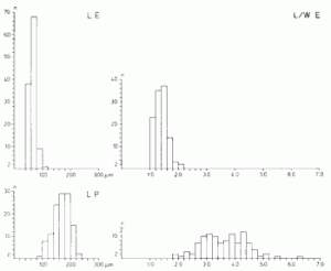
Analyser av längden och längd/bredd-kvoten hos pericysten och endocysten av olika arter visar att pericystens variation i allmänhet är större än endocystens. Endocysten har en mindre långsträckt form som är tämligen konstant inom en art (figur 2). Medelvärdet för längd/bredd-kvoten för endocysten varierar för de arter jag undersökt mellan 0.8 och 1.4 med medianvärdet = 1.0, medan medelvärdet för längd/bredd-kvoten för pericysten varierar mellan 1.3 och 4.5, med medianvärdet = 2.1

figur 2


Figure 2. Peridinioid dinoflagellate Deflandrea diebeli. Distribution of length (L) and length/width ratio (L/W) of endocyst (E) and pericycst (P). n = number of specimens.


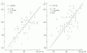
En ändring av cystans form mellan olika stratigrafiska nivåer kan visa på ett utvecklingsförlopp som orsakar en morfologisk förändring av fenotypen (figur 3).

 figur 3


Figure 3. Peridinioid dinoflagellate Paleoperidinium pyrophorum. Scatter diagrams with regression line showing cyst length (L) to cyst width (W) in two different samples. The slope (m), correlation coefficient (r), and number of specimens (n) are recorded on the figure. The correlation betweenL and W is statistically significant on the 0.1% level


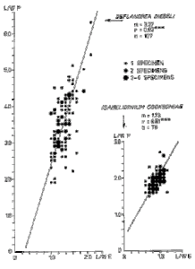
Förhållandet mellan formen hos pericysten och endocysten är relativt konstant inom ett släkte men varierar mellan olika släkten (figur 4 och figur 5).

 figur 4


Figure 4. Peridinioid dinoflagellates Deflandrea diebeli and Isabelidinium cooksoniae. Scatter diagrams with regression line showing length/width ratio of pericysts (L/W P) to length/width ratio of endocysts /L/W E). The slope (m), correlation coefficient (r), and number of specimens (n) are recorded on the figure. The correlation between L/W P and L/W E is statistically significant on the 0.1 % level.

 figur 5_750

Figure 5. Regression lines based on reduced majoraxis in scatter diagrams showing length/width ratio of pericysts (P) to length/width ratio of endocysts (E) for species of peridinioid dinoflagellates of genera Deflandrea, Isabelidinium, Subtilisphaera, and Trithyrodinium from the Trelleborg boring core T-1 (southern Sweden). The slope (m), correlation coefficient (r) and its significance level (* = 5 %, ** = 1 %, *** = 0.1 %), and number of specimens (n) are recorded on the figure.


Den begränsade variationen av storleken och formen hos endocysten av de studerade dinoflagellaterna visar att den inre väggen utvecklades under genetisk kontroll inuti en theca. Pericysten visar en vid morfologisk variation som kan vara resultat av påverkan av miljöfaktorer under den yttre väggens utveckling.

De båda släktena Isabelidinium och Eurydinium skiljs genom formen hos endocysten. I Isabelidinium skall längden av endocysten vara lika stor som eller mindre än bredden (L/W<1.0) medan i Eurydinium skall längden av endocysten vara större än bredden (L/W>1.0).

 figur 6


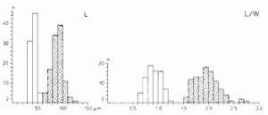
Figure 6. Peridinioid dinoflagellate Isabelidinium cooksoniae. Distribution of length (L) and length/width ratio (L/W) of endocyst (blank) and pericycst (dotted). n = number of specimens.


I det material jag studerat varierar längd/bredd-kvoten hos en art mellan 0.6 och 1.2. Kvoten är normalfördelad och har medelvärdet = 0.9 (figur 6). Detta visar att det inte finns någon anledning att särskilja dessa två släkten med endocystens form som kriterium.


Referens

Lindgren, S., 1984.
Acid resistant peridinioid dinoflagellates from the Maastrichtian of Trelleborg, southern Sweden. (Summary in Russian.) Stockholm Contrib. Geol., 39(6): 145—201. Stockholm. ISSN 0585-3532. ISBN 91-22-00519-6. — Buy at the lowest prices among books in Sweden.

Leiospheres

24 July, 2009
 
Leiosphere is a common name without taxonomic implications for thin-walled smooth or slightly ornamented spherical microalgae. The botanical affinities of leiospheres are unknown.Simple, thin-walled spheres, like Leiosphaeridia, may develop in a variety of algal taxa. When found as fossil remains, these spheres may appear morphologically undistinguishable, despite they are naturally unrelated.
Many acid resistant spherical microfossils have been recorded in sediments ranging from the early pre-Cambrian to Recent. They are widespread in pre-Cambrian and Paleozoic deposits.


The taxonomic classification is hazardous and complex in this group where the only features for classification are wall structure, ornamentation, and opening mechanisms. These features are not sufficient for a classification to modern algal taxa above the rank of species—not even at the rank of class.

The importance assigned to morphological characters for classification at the rank of genus or species appears merely to be a question of personal opinion. For modern algae neither shape or ornamentation, nor apertures are relevant taxonomic characters at the rank of genus.

Leiospheres have generally been considered as resting cysts and their aperture (pylome) has been interpreted as an excystment opening. However, they may also represent vegetative stages of the algal life cycle. There may be no possibility of discriminating between vegetative and resting stages (cysts) in fossil material.


    FORWARD TO


Morphology and taxonomy
Botanical affinities
Diagnostic features in modern algae
Morphometry of modern and fossil algae
Genus Leiosphaeridia
Distribution of Leiosphaeridia


References

Lindgren, S., 1981.
Remarks on the taxonomy, botanical affinities, and distribution of leiospheres. (Summary in Russian) Stockholm Contrib. Geol., 38(1): 1—20. Stockholm. ISBN 91-22-00500-5. ISSN 0585-3532. — Buy at the lowest prices among books in Sweden.
Lindgren, S., 1982.
Taxonomic review of Leiosphaeridia from the Mesozoic and Tertiary. Stockholm Contrib. Geol., 38 (2): 21—33. Stockholm. ISBN 91-22-00502-1. ISSN 0585-3532. — Buy at the lowest prices among books in Sweden.
Lindgren, S., 1982.
Algal coenobia and leiospheres from the Upper Riphean of the Turukhansk region, eastern Siberia. Stockholm Contrib. Geol., 38 (3): 35—45. Stockholm. ISBN 91-22-00504-8. ISSN 0585-3532. — Buy at the lowest prices among books in Sweden.
Lindgren, S., 1982.
A new taxon of Leiosphaeridia (algae) from Upper Cretaceous clays, southern Sweden. Stockholm Contrib. Geol., 37 (11): 139—143. Stockholm. ISBN 91-22-00487-4. ISSN 0585-3532.
Lindgren, S., 1984.
A new taxon of Leiosphaeridia (algae) from Upper Cretaceous clays, southern Sweden. Stockholm Contrib. Geol., 39 (5): 139—144. Stockholm. ISBN 91-22-00517-x. ISSN 0585-3532. — Buy at the lowest prices among books in Sweden.


Web Links

Steiner, M., 1997. Chuaria circularis Walcott 1899 — “megasphaeromorph acritarch” or prokaryotic colony? Acta Universitatis Carolinae, Geologica, 40: 645—665. Prague.

Halosphaera, a small planktonic flagellate and a large planktonic cyst; by Charles J. O’Kelly, Protist Image Database

Introduction to the Micromonadophyceae; from University of California, Berkeley.

Non-Marine Palynomorphs from the Middle Cambrian Bright Angel Shale, Grand Canyon USA. Paul K. Strother, Weston Observatory, Boston College.

Terrestrial Organic Remains from Lower Paleozoic Paralic Sequences. Paleobotany Laboratory at Weston Observatory. In Research Projects.

Palynology at the Hole. Rob Fensome and Andrew MacRae, Canadian Association of Palynologists.


Morphology and taxonomy of Leiospheres

24 July, 2009


Many acid resistant spherical microfossils have been recorded in sediments ranging from the early pre-Cambrian to Recent.

Leiosphere is a common name without taxonomic implications for thin-walled smooth or slightly ornamented spherical microalgae. The botanical affinities of leiospheres are unknown.

Simple, thin-walled spheres, like Leiosphaeridia, may develop in a variety of algal taxa. When found as fossil remains, these spheres may appear morphologically undistinguishable, despite they are naturally unrelated.

No features used for classification of modern algae—even at the rank of class—are preserved in fossil leiospheres. It is evidently impossible also to discriminate between different morphological appearances due to ontogenetic variability and environmental modifications. Consequently the leiospheres must be arranged in an artificial system without biological implications, but as far as possible according to biological principles.

Above the rank of genus leiospheres have been placed in morphologic groupings that are outside the sequence of ranks of taxa treated under the International Code of Botanical Nomenclature. They are not determined by means of nomenclatural types and not based upon priority of publication. Thus, all such groupings are equally applicable. The name acritarch meaning “of uncertain origin” was used by Evitt in 1963.

As morphologic groupings above the rank of genus in a completely artificial scheme of classification do not reflect any natural or genetical relations, the genera may better be arranged and discussed simply in alphabetical order. This may be particularly relevant because the algal cell may display a variability of shape during the life cycle.

The taxonomic classification is hazardous and complex in this group where the only features for classification are wall structure, ornamentation, and opening mechanisms. These features are not sufficient for a classification to modern algal taxa above the rank of species—not even at the rank of class.

Ornamentation is the main taxonomic character in several genera in this complex group, and has been used also as a diagnostic character at the rank of genus. However, in classification of modern algae it is relevant only at the rank of species. Even size has been used in some taxa as the only character of diagnostic value.

The importance assigned to morphological characters for classification at the rank of genus or species appears merely to be a question of personal opinion. For modern algae neither shape or ornamentation, nor apertures are relevant taxonomic characters at the rank of genus.

The wide size range from 8 µm to 440 µm in Leiosphaeridia indicates that different natural groups are included in the genus, but in comparison with modern algae it does not exclude definitely, however, that it is a natural genus.

In the leiospheres there are many badly defined and partly superfluous taxa. Several species are synonymous because they have been assigned to different genera which are synonymous. Species overlap because of different interpretations of ranges of diagnostic characters among investigators. Inadequate illustrations and insufficient diagnoses have made identification difficult. Accidental and diagenetic features have been interpreted as taxonomically valid.

However, some forms of leiospheres are morphologically well defined and have a restricted variability, at least in the same stage of the life cycle. They are distinct form species, no matter the name and circumscription of the genus to which they may belong. These forms are considered useful for biostratigraphic correlations and age determinations, specially in the pre-Cambrian and lower Palaeozoic.

    FORWARD TO


Botanical affinities
Diagnostic features in modern algae
Morphometry of modern and fossil algae
Genus Leiosphaeridia
Distribution of Leiosphaeridia


References

Lindgren, S., 1981.
Remarks on the taxonomy, botanical affinities, and distribution of leiospheres. (Summary in Russian) Stockholm Contrib. Geol., 38(1): 1—20. Stockholm. ISBN 91-22-00500-5. ISSN 0585-3532. — Buy at the lowest prices among books in Sweden.
Lindgren, S., 1982.
Taxonomic review of Leiosphaeridia from the Mesozoic and Tertiary. Stockholm Contrib. Geol., 38 (2): 21—33. Stockholm. ISBN 91-22-00502-1. ISSN 0585-3532. — Buy at the lowest prices among books in Sweden.
Lindgren, S., 1982.
Algal coenobia and leiospheres from the Upper Riphean of the Turukhansk region, eastern Siberia. Stockholm Contrib. Geol., 38 (3): 35—45. Stockholm. ISBN 91-22-00504-8. ISSN 0585-3532. — Buy at the lowest prices among books in Sweden.
Lindgren, S., 1982.
A new taxon of Leiosphaeridia (algae) from Upper Cretaceous clays, southern Sweden. Stockholm Contrib. Geol., 37 (11): 139—143. Stockholm. ISBN 91-22-00487-4. ISSN 0585-3532.
Lindgren, S., 1984.
A new taxon of Leiosphaeridia (algae) from Upper Cretaceous clays, southern Sweden. Stockholm Contrib. Geol., 39 (5): 139—144. Stockholm. ISBN 91-22-00517-x. ISSN 0585-3532. — Buy at the lowest prices among books in Sweden.


Web Link

Are the green algae (phylum Viridiplantae) two billion years old? by Bernard Teyssèdre, Paris. Notebooks on Geology, Article 2006/03 (CG2006_A03), September 19, 2006


Botanical affinities of Leiospheres

24 July, 2009


Leiospheres have generally been considered as resting cysts and their aperture (pylome) has been interpreted as an excystment opening. However, they may also represent vegetative stages of the algal life cycle, as in modern acid resistant microalgae apertures similar to pylomes may develop also in vegetative stages, e.g. as flagellar pores.

There may be no possibility of discriminating between vegetative and resting stages (cysts) in fossil material. The acid resistance and the possibility to be fossilized are widespread in the algae and occur variably in all their life stages, in vegetative cells and also in resting cysts.


Apertures

Apertures have a restricted taxonomic significance and they are not useful for the determination of life stages. Vegetative stages of algae and also encysted resting stages are likely to be preserved as fossils. Apertures on vegetative cells, however, cannot always be definitely discriminated from excystment openings. The pylome may be a flagellar pore similar to that of certain modern algae (e.g. Trachelomonas, see below).

If the fossils are of flagellate affinity, every one should have an aperture in its vegetative stage. Forms without apertures may be cysts of the same or different species, but also vegetative stages of unflagelled taxa. When the aperture is an excystment feature, it is present only during one life stage. Cells without apertures then may be closed cysts, but also vegetative stages of other than flagellate affinity. Cells with apertures may be mature cysts, but also vegetative stages of flagellate affinity.

In the original diagnosis of Leiosphaeridia (Eisenack 1958) pylome (aperture) is said to be present. In the following original description is remarked that pylomes are common in some species but rare or absent in others. This variation is proposed to be depending either on specific characters or on the stage of the life cycle of the organisms at the time when they were buried. Microfossils with pylomes were interpreted as obviously abandoned cells in their final life stage, and the function of the opening, was without doubts considered to be making it possible for the protoplast to extrude. Consequently pylomes are not necessarily present on every fossil specimen of a taxon, and their occurrence has no taxonomic value. Only their structure may be a feature important for the classification, possibly limited to the rank of species.

In some taxa a pylome is always present, in others pylomes are never or only occasionally observed with certainty. In “thousands of specimens” of the original material of Leiosphaeridia plicata and L. ralla there was no pylome (Felix 1965), and in 3324 specimens of L. wenlockia there were “one or two” with pylome (Downie 1959).

Generally, leiospheres have been considered as cysts having pylomes and ruptures as excystment features. However, since excystment is a genetically controlled characteristic process that is constant within a taxon, pylomes and ruptures cannot exist both as excystment features in the same species.


Environmental modifications

In the fossil record most often it is impossible to elucidate if variability is due to environmentally controlled factors or to different stages of the algal life cycle. Thus, different fossil genera from distant deposits may be only modifications of the same natural species.

Cells that do not differentiate hard parts do not always obtain identical morphological features when they are developed separately in different environments. Except this environmentally controlled morphogenesis the different life stages of the algal cell may generate morphologic variability. Further, in restricted environments disastrous events may occur which may often generate radical changes in the metabolism and morphological appearance of the cells.

A satisfactory classification of modern algae must pay sufficient attention to the morphological variability and life history of each taxon. Many species first described on the basis of specimens representing only one stage of their life cycles were later defined as life stages of other species.


Coenobia

I have identified fossil algal coenobia in a sample from the Upper Riphean Mirojedikha Beds in the Turukhansk region, eastern Siberia. The fossils I investigated derive from the Mirojedikha River and were extracted from a sample from the Upper Riphean Mirojedikha Beds.

The Mirojedikha Beds are about 200 m thick shallow water sediments consisting of dolostones, limestones, and shales. The present sample was collected in l967 by my friend, the Late Professor Boris V. Timofeev at the type locality at the Mirojedikha River, four kilometers from its confluence with the Yenisey River, 30 kilometers north of the town of Turukhansk. The sample is a dark-grey, clayey shale which derives from the lower part of the Mirojedikha Beds.

Vesicles of coenobia and solitary leiospheres do not display morphologic differences. The dimensions of vesicles of coenobia correspond to those of larger specimens in the bimodal size distribution of solitary leiospheres from the same sample.

Algal coenobia are sparsely reported as fossil findings. From the Cretaceous-Tertiary they are described as the genus Palambages. The first findings of coenobia in the Palaeozoic were reported from the Silurian of Libya in 1962. There were not any records from the Precambrian until my findings in 1982.

Faulty or rough processing may be the reason for the scantiness of reports of fossil colonies. Maybe most samples yield colonies rather than single specimens if they are treated with gentle care. Clusters, flakes, and different kinds of agglomerations of uniform fossil microalgae have been reported as colonies though they do not strictly represent colonies.

A coenobium is a definitely integrated colony composed of a certain number of cells which are determined during its early development; after the embryonic phase cell-division does not occur until the following reproductive phase. In coenobia the joints and sheaths are usually resistant and likely to be preserved as fossils. However, unicell production and colony formation are influenced by environmentaly controlled factors.

In the algae there is a parallel development from simple to more elaborate forms so that practically all types of cellular and colonial organizations have their counterparts in different classes. The characters necessary for discrimination between classes are not preserved as fossils. Consequently, relating fossil coenobia to modern algal taxa is impossible.

I referred both coenobial and solitary forms to a species with the new combination name Leiosphaeridia asperata (Naumova) Lindgren. Kildinella hyperboreica is not the correct name of this taxon. Protoleiosphaeridium, Kildinella, and Polyedrosphaeridium are congeneric with Leiosphaeridia, as well as the not validly published genera Leiopsophosphaera, Macroptycha, and Scaphita.

BACK TO


Morphology and taxonomy

  FORWARD TO


Diagnostic features in modern algae
Morphometry of modern and fossil algae
Genus Leiosphaeridia
Distribution of Leiosphaeridia


References

Downie, C., 1959.
Hystrichospheresfrom the Silurian Wenlock Shale of England. Paleontology, 2: 26—71. London.
Eisenack, A., 1958.
Tasmanites Newton 1875 und Leiosphaeridia n.g. als Gattungen der Hystrichosphaeridea. Palaeontographica Abt.A, 110: 1—19. Stuttgart.
Felix, C.J., 1965.
Neogene Tasmanites and leiosphere from southern Louisiana, U.S.A Palaeontology, 8: 16—26. London.
Lindgren, S., 1981.
Remarks on the taxonomy, botanical affinities, and distribution of leiospheres. (Summary in Russian) Stockholm Contrib. Geol., 38(1): 1—20. Stockholm. ISBN 91-22-00500-5. ISSN 0585-3532. — Buy at the lowest prices among books in Sweden.
Lindgren, S., 1982.
Taxonomic review of Leiosphaeridia from the Mesozoic and Tertiary. Stockholm Contrib. Geol., 38 (2): 21—33. Stockholm. ISBN 91-22-00502-1. ISSN 0585-3532. — Buy at the lowest prices among books in Sweden.
Lindgren, S., 1982.
Algal coenobia and leiospheres from the Upper Riphean of the Turukhansk region, eastern Siberia. Stockholm Contrib. Geol., 38 (3): 35—45. Stockholm. ISBN 91-22-00504-8. ISSN 0585-3532. — Buy at the lowest prices among books in Sweden.
Lindgren, S., 1982.
A new taxon of Leiosphaeridia (algae) from Upper Cretaceous clays, southern Sweden. Stockholm Contrib. Geol., 37 (11): 139—143. Stockholm. ISBN 91-22-00487-4. ISSN 0585-3532.
Lindgren, S., 1984.
A new taxon of Leiosphaeridia (algae) from Upper Cretaceous clays, southern Sweden. Stockholm Contrib. Geol., 39 (5): 139—144. Stockholm. ISBN 91-22-00517-x. ISSN 0585-3532. — Buy at the lowest prices among books in Sweden.


Diagnostic features in modern algae comparable to Leiosphers

24 July, 2009


Chlorococcales

The features considered to be of greatest taxonomic importance at the rank of genus in the modern Chlorococcales are chromatophores, pyrenoids, and zoospores. None of these are preserved in fossils. Modifications due to environmental influences cannot always be discriminated from hereditary variability by microscopic analyses alone. In preserved modern material it is impossible to extract the various stages necessary for distinguishing genera and species. That is also a major difficulty in mixed cultures of living material—and of course in fossil material. Thus, cultivation is the essential method for taxonomic classification of many unicellular modern algae.

The fossil leiospheres lack all the characters mentioned above, which are necessary for their classification in modern algal taxa—even at the rank of class. It is evidently impossible also to discriminate between different morphological appearances due to ontogenetic variability and environmental modifications. Consequently the leiospheres must be arranged in an artificial system without biological implications, but as far as possible according to biological principles.

Cysts are metabolically dormant cells with a considerably higher resistance than vegetative cells to detrimental physical conditions, other than heat. Desiccation resistance may be a survival attribute in nature, and this property seems to be related to the outer cell wall.

The size is not a sole diagnostic character. Anyhow related modern taxa (genus, species) seem to have a limited size range so measurements may be important features for determination of modern algae. I have compared the size distribution in Leiosphaeridia to vegetative stages of modern unicellular Chlorococcales.

Cyanophyceae

In the Cyanophyceae the normal reproductive cell, the akinete, is a spherical or cylindrical cell with a smooth or granulated surface. It is characterized by a considerable increase in size compared with the vegetative cell, and before maturation it will expand still more. The ultrastructure is similar to that of the vegetative cell, but with a new outer wall added. Thus the akinete can be regarded as an enlarged vegetative cell enclosed by a thick outer envelope.

Akinete-like cells with thickened walls develop also in the Chlorophyceae, but conditions associated with the production of this stage were not sufficiently investigated.

Akinete germination occurs in different ways. Frequently it generates the rupturing of the outer wall. The excystment mechanism is a true taxonomical feature. According to genetical control, some taxa will dehisce by an aperture (pylome), others by a rupture. However, this is recognizable only in certain stages of their life cycles.


Modern algae Trachelomonas

My laboratory investigations of the modern euglenophycean algae Trachelomonas volvocina showed that the lorica of these specimens is acid resistant and that after treatment in acids their flagellar pore is similar to the pylome of leiospheres. The living cells are spherical but they collapse during the acetolyzis process and form folds similar to those said to characterize the fossil genus Kildinella (=Leiosphaeridia).

Trachelomonas is characterized by a lorica that is often iron impregnated, with an apically located flagellar pore, and a very long flagellum. The species are discriminated by shape, ornamentation, processes, and flagellar pore features. A series of 447 illustrations (Conrad & van Meel 1952) of Trachelomonas species shows that shape, ornamentation, processes, and aperture characteristics are not significant taxonomic features at the rank of genus.

About 250 species of Trachelomonas are known. The cells show a considerable variability in size and shape, which makes the taxonomy of the group complex. This variability is also due to different stages of the life history of the organisms and to environmentally controlled morphogenesis. In acidic water some species may have a hyaline, smooth lorica, whereas the same species in alkaline water have thickened lorica impregnated with iron- and manganese-compounds. Exposed to unfavourable environmental conditions some species of Trachelomonas will develop into round, thick-walled resting cells that may be difficult to discriminate from specimens of other genera, even of different classes, e.g. the chlorophycean genus Chlamydomonas.

Specimens of Trachelomonas shown on the internet.

BACK TO


Morphology and taxonomy
Botanical affinities

  FORWARD TO


Morphometry of modern and fossil algae
Genus Leiosphaeridia
Distribution of Leiosphaeridia


References

Conrad, W. & van Meel, L., 1952.
Matériaux pour une monographie de Trachelomonas Ehrenberg, C., 1834, Strombomonas Deflandre, G., 1930 et Euglena Ehrenberg, C., 1832, Genres d’Euglénacées. Mém. Inst. R. Sci. Nat. Belg., 124: 1—176, pls. 1—13.
Lindgren, S., 1981.
Remarks on the taxonomy, botanical affinities, and distribution of leiospheres. (Summary in Russian) Stockholm Contrib. Geol., 38(1): 1—20. Stockholm. ISBN 91-22-00500-5. ISSN 0585-3532. — Buy at the lowest prices among books in Sweden.
Lindgren, S., 1982.
Taxonomic review of Leiosphaeridia from the Mesozoic and Tertiary. Stockholm Contrib. Geol., 38 (2): 21—33. Stockholm. ISBN 91-22-00502-1. ISSN 0585-3532. — Buy at the lowest prices among books in Sweden.
Lindgren, S., 1982.
Algal coenobia and leiospheres from the Upper Riphean of the Turukhansk region, eastern Siberia. Stockholm Contrib. Geol., 38 (3): 35—45. Stockholm. ISBN 91-22-00504-8. ISSN 0585-3532. — Buy at the lowest prices among books in Sweden.
Lindgren, S., 1982.
A new taxon of Leiosphaeridia (algae) from Upper Cretaceous clays, southern Sweden. Stockholm Contrib. Geol., 37 (11): 139—143. Stockholm. ISBN 91-22-00487-4. ISSN 0585-3532.
Lindgren, S., 1984.
A new taxon of Leiosphaeridia (algae) from Upper Cretaceous clays, southern Sweden. Stockholm Contrib. Geol., 39 (5): 139—144. Stockholm. ISBN 91-22-00517-x. ISSN 0585-3532. — Buy at the lowest prices among books in Sweden.

Morphometry of modern Chlorococcales and fossil Leiosphaeridia (algae)

24 July, 2009


The size is not a sole diagnostic character. Anyhow related modern taxa of the rank of genus as well as species seem to have a limited size range and measurements may be important features for determination of modern algae.

The size distribution in Leiosphaeridia may be compared to vegetative stages of modern unicellular Chlorococcales. The knowledge of size relations between vegetative cells and resting cysts is too fragmentary for comparisons.

For comparisons of dimensions among taxa a “size difference” value (SD) may be calculated as the size range value in per cent of the minimum size value (Lindgren 1981).

size


Figure 1
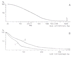
A. Cumulative diagram showing size differences in 49 genera of the Chlorococcales. Size difference of Leiosphaeridia is indicated by an L.
B. Cumulative diagram showing size differences in 257 species of the Chlorococcales (curve C), and in 55 species of Leiosphaeridia (curve L).
The size difference is calculated as the size range value in per cent of the minimum value. (From Stockholm Contrib. Geol., 38(1): 8, figure 1)


In 49 genera of the Chlorococcales tabulated from Philipose (1967) the size difference varies between SD = 4 and SD = 5400, with a median value of SD = 250 (fig. 1A). Only in six genera the size difference exceeds SD = 1000, and only in two it exceeds SD = 4000. Specimens of Ankistrodesmus Corda 1838 emend. Ralfs 1848 (seven species) range from 3 µm to 165 µm (SD = 5400), and specimens of Chlorococcum Fries 1820 (three species) range from 2 µm to 109 µm (SD = 5350).

In Leiosphaeridia the size range is recorded as 8—440 µm. The size difference is SD = 5400.

Thus the size difference for genus Leiosphaeridia is extremely high, but not quite impossible for a natural taxon. In described species of Leiosphaeridia, however, the size range is far more restricted than in modern Chlorococcales (fig 1B).

In 257 species of the Chlorococcales, also tabulated from Philipose (1967), the size difference in vegetative cells ranges between SD = 4 and SD = 1163, with a median value of SD = 100. 13.6 % of the difference values exceed SD = 200 and 4.7 exceed SD = 350.

In 55 species of Leiosphaeridia (including Protoleiosphaeridium) the size difference ranges from SD = 3 to SD = 203, with a median value of SD = 58.

The values of the size differences are summarized in table I


Table I
Size differences in modern Chlorococcales and fossil Leiosphaeridia


Number 


Size difference 


of taxa 


Minimum 


Maximum 


Median 


Mean 


Genera             
Chlorococcales

49


4


5400


250


557


Leiosphaeridia 

1


5400


Species             
Chlorococcales

257


4


1163


100


128


Leiosphaeridia 

55


3


203


58


62


 The size difference is calculated as the size range value in per cent of the minimum value.


BACK TO


Morphology and taxonomy
Botanical affinities
Diagnostic features in modern algae

  FORWARD TO


Genus Leiosphaeridia
Distribution of Leiosphaeridia


References

Lindgren, S., 1981.
Remarks on the taxonomy, botanical affinities, and distribution of leiospheres. (Summary in Russian) Stockholm Contrib. Geol., 38(1): 1—20. Stockholm. ISBN 91-22-00500-5. ISSN 0585-3532. — Buy at the lowest prices among books in Sweden.
Philipose, M.T., 1967.
Chlorococcales. Indian Counc. Agric. Res., New Delhi. 365 p.


Web Link

Steiner, M., 1997. Chuaria circularis Walcott 1899 — “megasphaeromorph acritarch” or prokaryotic colony? Acta Universitatis Carolinae, Geologica, 40: 645—665. Prague.


Genus Leiosphaeridia

24 July, 2009


The taxonomic classification of spherical microalgae is very complex, since:

  • it is impossible to refer these forms to modern algal taxa because of the lack of relevant characters for classification
  • vegetative cells and resting stages (cysts) cannot be discriminated with respect to only fossilized parts;
  • cell shape and ornamentation may vary between different stages of the algal life cycle and can be modificatively affected by environmental factors, such as nutrition and temperature
  • excystment features (pylomes, ruptures) are not constant in the algal life cycle, and excystment apertures (pylomes) cannot be discriminated always with certainty from apertures in vegetative stages, e.g. flagellar pores.

Leiosphaeridia (Eisenack 1958) comprises acid resistant, spherical to ovoidal microfossils without processes, without divisions into fields, and without traverse and longitudinal furrows or girdles. The cell wall is thin and without tubes, the surface is smooth or with slight ornamentation. An aperture (pylome) may be present, and has been considered as an excystment mechanism. Other methods of dehiscence are also recorded, e.g. by a split.

The original diagnosis by Eisenack in 1958 was slightly emended by Downie and Sarjeant in 1963 to include slight ornamentation and to exclude reference to colour. However, the view on color was taken earlier, and published by Eisenack in 1962.

Leiosphaeridia was established to accommodate leiospheres not attributable to Tasmanites (Newton 1875) because the nomenclatural type of Leiosphaera—a genus introduced by Eisenack in 1938—proved to be conspecific with a species of Tasmanites.


Synonymy

  • Kildinella, Lophosphaeridium, and Protoleiosphaeridium do not display any differences from the diagnosis of Leiosphaeridia. Also Leiopsophosphaera and Uniporata seem to be congeneric with Leiosphaeridia.
  • Leiosphaeridium is an illegitimate name. Macroptycha and Scaphita are not validly published.
  • Campenia and Lancettopsis may be congeneric with Leiosphaeridia, but are not examined.

Kildinella (Shepeleva & Timofeev ex Timofeev) comprises according to the protologue smooth or shagreen spherical microfossils, ranging from 15 to 70 µm in diameter, with clearly delimited folds. They differed from Leiosphaeridia specimens only in being smaller and having denser membranes.

The folds evidently were generated by compression and the restricted size range is of no taxonomic value. When I examined the original material of Kildinella, I could not find any distinct features different from the diagnosis of Leiosphaeridia.

Kildinella was established by Shepeleva & Timofeev in 1963, but no species was described and no nomenclatural type was indicated. Thus the name was not validly published until Timofeev in 1966 described Kildinella hyperboreica and designated it as the type species.

Only a few species of Kildinella have been described. My friend, the Late Professor Boris V. Timofeev showed to me in Leningrad 1979 that they are essential parts of biostratigraphically useful microalgal assemblages identified in the pre-Cambrian of the Soviet Union.

Lophosphaeridium (Timofeev ex Downie) differs from Leiosphaeridia only in having a tuberculose ornamental sculpture. The difference is diffuse and a more or less developed ornamentation is the only feature for the generic classification. When I examined the original material of Lophosphaeridium, I could not find any distinct features different from the diagnosis of Leiosphaeridia. However, I regarded more extensive analyses required for establishing the taxonomic relations.

Lophosphaeridium was introduced by Timofeev in 1959 but as no type species was selected it was not valid until Downie designated the nomenclatural type in 1963.

Protoleiosphaeridium comprises small leiospheres (less than 50 µm in diameter) with smooth or shagreen surface, and ranges completely within the diagnosis of Leiosphaeridia. This genus was established by Timofeev in 1959, and became valid by designation of a lectotype in 1960. The circumscription of the genus was slightly expanded by Staplin in 1961 as to include all types of minor overall ornamentation. Leiosphaeridia and Protoleiosphaeridium were treated as congeneric by Downie & Sarjeant in 1963.

Leiopsophosphaera (Naumova) comprises large cells with smooth or shagreen, pitted surface. Dr Pykhova told me in 1978 that Leiopsophosphaera differs from Leiosphaeridia and Uniporata only in the sculptural ornamentation. This Proterozoic and Palaeozoic genus seems to range within the diagnosis of Leiosphaeridia.

A more detailed investigation of the taxonomy should indude a close examination of the original material if it is preserved. Studying that material was not possible for me, nor was I able to find Naumova’s paper from 1960 with the original diagnosis. Leiopsophosphaera and Leiosphaeridia were treated as congeneric by Yin & Li in 1978.

Uniporata (Naumova in Pykhova) comprises cells with ornamented surface and a large pylome. Dr Pykhova told me in 1978 that Uniporata differs from Leiosphaeridia only in having a large pylome. The type species indicated in the protologue of Uniporata is a nomen nudum and thus the name is not validly published.

The Proterozoic and Palaeozoic genus Uniporata was established by Naumova in Pykhova 1969. It seems to range within the diagnosis of Leiosphaeridia. A more detailed investigation of the taxonomy should indude a close examination of the original material if it is preserved. Studying that material was not possible for me.

Leiosphaeridium was proposed by Timofeev in 1959 as an amendment of Eisenack’s Leiosphaera. The original spelling of a name is to be retained and Leiosphaeridium is not to be considered an orthographic variant of Leiosphaera. As the name is nomenclaturally superfluous, it is illegitimate.

Staplin emended Timofeev’s diagnosis of Leiosphaeridium and regarded the name “as a new generic taxon” with L. eisenackii (Timofeev) as a new type species. Based on a different type the name is a later homonym and thus illegitimate.

The transference to Leiosphaeridia of Leiosphaeridium eisenackii made by Downie & Sarjeant in 1963 is not valid since it is done without clear references to the basionym.

Macroptycha and Scaphita are names used by Timofeev in 1973 for boat-shaped forms with large longitudinal folds or without folds, respectively. However, both names were not validly published. The folds were interpreted as prolonged chambers, but evidently represent compressional features. Before 1973 forms identical with Macroptycha and Scaphita were referred to Leiosphaeridia by Timofeev and others.

Campenia, described by Mädler in 1963, differs from Leiosphaeridia in possessing an elliptical slit that is supposed to be homologous with the pylome. The diagnosis of Leiosphaeridia, however, does not exclude opening by a slit. Lancettopsis—also described by Mädler in 1963—comprises folded and rolled up vesicles similar to Scaphita. Analyses of the original material is required for establishing the taxonomic relations.


Complex classification

To illustrate the complex leiosphere classification two species may be mentioned.

Protoleiosphaeridium papillatum was described by Staplin in 1961. Two years later—in 1963—it was transferred (but not validly) to Leiosphaeridia by Downie & Sarjeant, and further five years later—in 1968—to Lophosphaeridium by Martin.

Protoleiosphaeridium granulosum was also introduced by Staplin in 1961. It was in 1963 transferred by Downie & Sarjeant to Leiosphaeridia, and in the same year by the same Downie (without Sarjeant as coauthor) to Lophosphaeridium. Both these transferences are, however, contrary to the nomenclatural rules, and the valid publication of the name Leiosphaeridia granulosa was made by Pocock in 1972—but for a different taxon!


New species

I have described two new species of acid resistant spherical microalgae, referable to genus Leiosphaeridia, from the Upper Cretaceous of the province Scania (Skåne), southern Sweden.

Leiosphaeridia nelliae Lindgren 1982c is described from a Cenomanian clay deposit penetrated by a boring at Åhus in the Kristianstad area. This species is diagnosed by its small size and the lack of apertures or any obvious dehiscence mechanism. It resembles some Palaeozoic species, but all Mesozoic species with similar appearance are much larger.

Leiosphaeridia scanica Lindgren 1984 is described from the Campanian and Maastrichtian penetrated by a boring at Trelleborg. This species differs from other species of Leiosphaeridia in having a smooth vesicle and a constantly present pylome.

Leiosphaeridia asperata (Naumova) Lindgren is a new combination I established in my 1982b paper with Trachytriletus asperatus Naumova as basionym.

BACK TO


Morphology and taxonomy
Botanical affinities
Diagnostic features in modern algae
Morphometry of modern and fossil algae

  FORWARD TO


Distribution of Leiosphaeridia


References

Eisenack, A., 1958.
Tasmanites Newton 1875 und Leiosphaeridia n.g. als Gattungen der Hystrichosphaeridea. Palaeontographica Abt. A, 110: 1—19. Stuttgart.
Eisenack, A., 1962.
Einigen Bemerkungen zu neueren Arbeiten uber Hystrichosphären. Neues Jb. Geol. Paläont. Mh., 102: 92—101. Stuttgart.
Downie,C. & Sarjeant, W.A.S., 1963.
On the interpretatio and status of some hystrichospher genera. Palaeontology, 6: 83—96. London.
Lindgren, S., 1981.
Remarks on the taxonomy, botanical affinities, and distribution of leiospheres. (Summary in Russian) Stockholm Contrib. Geol., 38(1): 1—20. Stockholm. ISBN 91-22-00500-5. ISSN 0585-3532. — Buy at the lowest prices among books in Sweden.
Lindgren, S., 1982a.
Taxonomic review of Leiosphaeridia from the Mesozoic and Tertiary. Stockholm Contrib. Geol., 38 (2): 21—33. Stockholm. ISBN 91-22-00502-1. ISSN 0585-3532. — Buy at the lowest prices among books in Sweden.
Lindgren, S., 1982b.
Algal coenobia and leiospheres from the Upper Riphean of the Turukhansk region, eastern Siberia. Stockholm Contrib. Geol., 38 (3): 35—45. Stockholm. ISBN 91-22-00504-8. ISSN 0585-3532. — Buy at the lowest prices among books in Sweden.
Lindgren, S., 1982c.
A new taxon of Leiosphaeridia (algae) from Upper Cretaceous clays, southern Sweden. Stockholm Contrib. Geol., 37 (11): 139—143. Stockholm. ISBN 91-22-00487-4. ISSN 0585-3532.
Lindgren, S., 1984.
A new taxon of Leiosphaeridia (algae) from Upper Cretaceous clays, southern Sweden. Stockholm Contrib. Geol., 39 (5): 139—144. Stockholm. ISBN 91-22-00517-x. ISSN 0585-3532. — Buy at the lowest prices among books in Sweden.